


.webp)
In an effort to broaden students' understanding of the relationship between physics and the development of health technology, the Faculty of Medicine, Universitas Sumatera Utara (FK USU), held a guest lecture entitled “MRI as an Application of Quantum Physics in Radiodiagnostics”. The activity, held at the Hall of RSP Prof. dr. Chairuddin P. Lubis USU, featured Dr. rer. nat. Freddy Haryanto, S.Si., M.Si., as the speaker on Friday, September 26, 2025, at the Hall of RSP Prof. dr. Chairuddin P. Lubis USU.
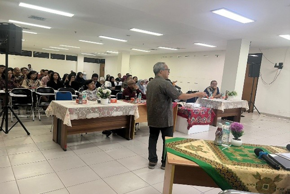
This guest lecture was part of an academic activity that provided students with an opportunity to gain a direct understanding of the application of physics concepts in medical technology. Through the presentation by a speaker with expertise in medical physics and medical imaging technology, students were invited to understand the relationship between fundamental physics theories and the technologies used in modern healthcare services.
In his presentation, Dr. rer. nat. Freddy Haryanto explained that Magnetic Resonance Imaging (MRI) is a medical imaging technology that utilizes principles of physics, particularly phenomena related to atomic nuclear spin, nuclear magnetic resonance, magnetic fields, and radio waves. Unlike examinations using X-rays or CT scans, MRI does not use ionizing radiation to produce images of body structures.
The discussion then focused on the relationship between MRI technology and quantum physics concepts. At the microscopic level, certain atomic nuclei, particularly hydrogen atoms that are abundant in the human body, have spin and magnetic momentum. When the body is placed in a strong magnetic field, some nuclear spins undergo a particular orientation. Applying radio waves at an appropriate frequency can cause resonance, while the resulting signals are then detected by the MRI system and processed using a computer to produce images of organs and body tissues.
Students were also introduced to several important concepts in MRI technology, including the main magnetic field, Larmor frequency, excitation using radiofrequency (RF) pulses, T1 and T2 relaxation, as well as image formation and reconstruction processes. Understanding these concepts provides an overview of how differences in tissue characteristics can be translated into visual information that supports the diagnostic process.
The material presented focused not only on the working principles of MRI but also demonstrated how phenomena at the atomic scale can be utilized to produce diagnostic information at the macroscopic scale. This demonstrates the relationship between fundamental physics, quantum physics, instrumentation technology, and medical science in the development of health technology.
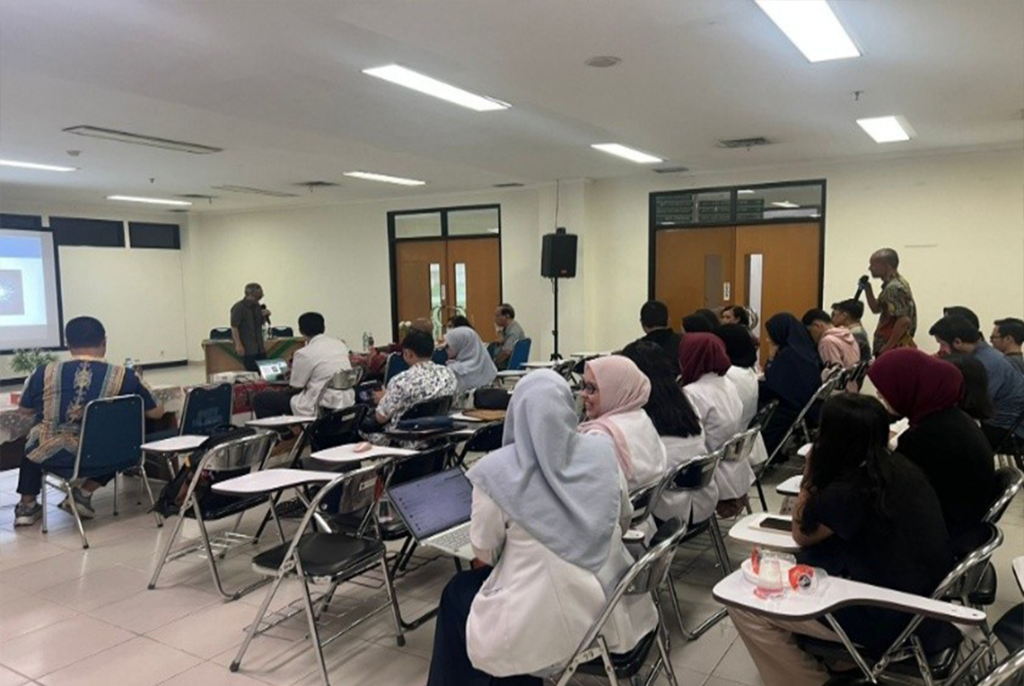
The activity was conducted interactively through a presentation and discussion session. Students were given opportunities to ask questions and discuss various aspects of the application of physics in radiodiagnostic technology. The interaction provided a space for students to connect the concepts they had learned theoretically with their application in medical technology used in the real world.
This activity also supports Sustainable Development Goal (SDG) 4: Quality Education through strengthening the learning process and developing student competencies. Exposure to interdisciplinary material provides students with opportunities to broaden their knowledge, develop critical thinking skills, and understand the relationship between fundamental sciences and their applications in healthcare. Such learning is part of efforts to develop human resources with knowledge and skills aligned with developments in science and technology.
In addition to the educational aspect, the discussion of MRI is also related to SDG 9: Industry, Innovation and Infrastructure. Medical imaging technology is one form of utilizing scientific and technological innovation to support healthcare services. Students' understanding of scientific principles and technological developments is expected to foster interest in healthcare innovation while broadening their perspectives on the role of technology in improving diagnostic capabilities.
Through the guest lecture “MRI as an Application of Quantum Physics in Radiodiagnostics”, students are expected not only to become familiar with MRI as a diagnostic tool but also to understand the scientific processes underlying its development and use. This knowledge can serve as a foundation for understanding developments in medical imaging technology while broadening students' perspectives on the application of physics in medicine.
The presence of Dr. rer. nat. Freddy Haryanto, S.Si., M.Si., provided an academic perspective that enriched the students' learning process, particularly in understanding how fundamental physics concepts can develop into technologies that provide benefits to healthcare. The activity is expected to encourage students to continue exploring developments in science, technology, and innovation that support modern healthcare services.